
Hip Pain & Tendon Injuries
Accurate diagnosis for hip pain using real-time ultrasound — so treatment matches the true source of pain.
Who This Is For
Hip pain is extremely common in:
- Middle-aged active adults
- Runners
- Lifters
- Women with lateral hip pain
- Individuals previously diagnosed with “bursitis” that never improved
- Patients who failed physical therapy alone
Many patients come in after months — or years — of persistent hip pain without clear answers.
The Hip Is a Complex Joint
Hip pain can originate from:
- Gluteus medius or minimus tendons
- Greater trochanteric bursa
- Rectus femoris tendon
- Hip joint (labrum or osteoarthritis)
- Proximal hamstring tendon
- IT band irritation
- Deep posterior hip structures
Symptoms overlap significantly.
Without imaging, lateral hip pain is often labeled “bursitis,” anterior pain is dismissed as tight hip flexors, and posterior pain is misattributed to the low back.
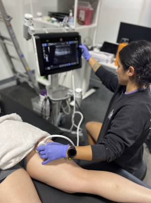
Ultrasound helps differentiate exactly where the pain and dysfunction are coming from — which completely changes treatment.
Using real-time musculoskeletal ultrasound, we evaluate:
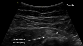
Lateral Hip Pathology
- Gluteus medius tendinopathy
- Gluteus minimus tendinopathy
- Partial gluteal tendon tears
- Greater trochanteric bursitis
- IT band-related irritation
Anterior Hip
- Rectus femoris tendinopathy
- Iliopsoas irritation
- Hip joint effusion
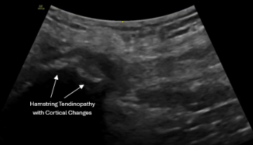
Posterior Hip
- Proximal hamstring tendinopathy
- Ischial region irritation
Intra-Articular Suspicion
- Labral-related pain
- Hip osteoarthritis
Dynamic ultrasound and side-to-side comparison allow us to correlate findings directly with your symptoms.

Every hip evaluation begins with diagnostic ultrasound paired with your functional complaints.
This allows us to:
- Differentiate tendon pathology from joint pathology
- Confirm partial tears
- Identify chronic degenerative changes
- Determine if injection is diagnostic or therapeutic
- Avoid unnecessary imaging when appropriate
Ultrasound also allows seamless transition into ultrasound-guided procedures during the same visit when indicated.
Precision-Guided Treatment
Treatment depends on:
- Ultrasound findings
- Symptom chronicity
- Activity demands
- Response to prior therapy
Corticosteroid Injections
Used selectively for:
- Diagnostic hip joint injections
- Significant inflammation
- Pain limiting rehabilitation
PRP (Platelet-Rich Plasma)
Frequently used for:
- Gluteus medius and minimus tendinopathy
- Partial gluteal tendon tears
- Proximal hamstring tendinopathy
- Hip osteoarthritis
- Labral-related pain
PRP is commonly performed under real-time ultrasound guidance for gluteal and hamstring tendons to ensure precise placement at the site of pathology.
All injections are image-guided to improve accuracy and safety.
Hip pain rarely exists in isolation.
Compensatory tension and fascial restriction often contribute to persistent symptoms.
We frequently incorporate:
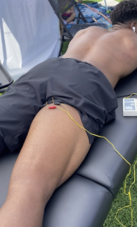
- Dry needling
- Cupping therapy
These are used to:
- Modulate pain
- Reduce surrounding muscular overactivity
- Improve fascial glide
- Support improved load tolerance
The goal is restoring functional movement — not simply reducing symptoms.
Ready to Get Clear Answers for Your Hip Pain?
If hip pain has not responded to rest or therapy — or you’ve been told it’s “just bursitis” without improvement — diagnostic ultrasound can identify the true source.