
Achilles, Ankle & Plantar Fascia Pain
Clear answers for heel, achilles, and ankle pain using diagnostic ultrasound and precision-guided treatment — so you can return to movement confidently.
Who This Is For
Achilles, ankle, and heel pain are extremely common in:
- Runners
- Lifters
- Jumping athletes
- Field sport athletes
- Active adults increasing training volume
- Adults with old ankle sprains that never fully healed
We frequently see patients with:
- Chronic Achilles pain lasting months or years
- Heel pain with first steps in the morning
- Pain with running or pushing off
- Recurrent ankle instability
- Persistent plantar fascia pain despite stretching
- Failed physical therapy alone
Many patients have already tried rest, rehab, inserts, or load modification — and want to know what tissue is actually injured.
Not All Heel Pain Is the Same
Pain in the back or bottom of the heel may come from:
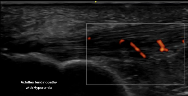
- Mid-substance Achilles tendinopathy
- Insertional Achilles tendinopathy
- Partial Achilles tears
- Retrocalcaneal bursitis
- Plantar fasciitis / plantar fasciopathy
- Partial plantar fascia tears
- Ankle joint or tendon pathology
- Scar tissue after previous sprains
Symptoms overlap. Without imaging, treatment often becomes generalized rather than targeted.
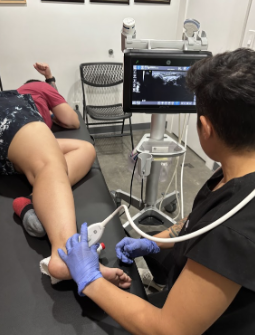
This is why we begin with diagnostic ultrasound.
Achilles, Ankle & Plantar Conditions We Diagnose
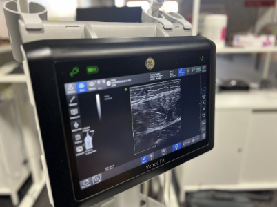
Using real-time musculoskeletal ultrasound, we evaluate:
Achilles Pathology
- Mid-portion tendinopathy
- Insertional tendinopathy
- Partial thickness tears
- Fat pad impingement
- Achilles bursitis
- Chronic degenerative tendon changes
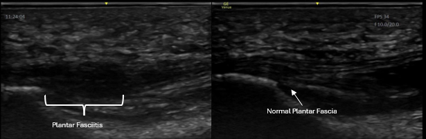
Plantar Fascia Pathology
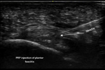
- Plantar fasciitis / fasciopathy
- Thickened or degenerative plantar fascia
- Partial plantar fascia tears
- Heel spur-related irritation
- Morton’s neuroma
Ankle Pathology
- Joint effusion
- Ankle instability
- Ligament partial or complete tears
- Peroneal tendon pathology
- Posterior tibial tendon dysfunction
- Post-sprain scar tissue
- Ganglion cysts
- Tibial nerve compression
Ultrasound allows us to:
- Directly visualize tissue structure
- Perform dynamic evaluation during movement
- Compare side to side
- Correlate imaging with symptoms in real time
You leave knowing exactly what is involved — not just a generic diagnosis of “heel pain.”
Diagnostic Clarity Changes the Plan
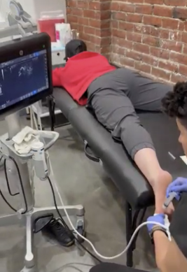
Every evaluation begins with ultrasound paired with your functional complaints.
This allows us to:
- Distinguish inflammation from chronic degeneration
- Identify partial tearing
- Avoid unnecessary imaging
- Determine whether injection-based treatment is appropriate
Ultrasound also allows seamless transition into ultrasound-guided procedures when needed.
Treatment is individualized based on:
- Ultrasound findings
- Duration of symptoms
- Severity of degeneration
- Training demands and goals
Corticosteroid Injections
Used selectively for:
- Retrocalcaneal bursitis
- Tenosynovitis
- Morton’s neuroma’s
- Fat pad impingement
- Severe inflammatory cases
- Pain significantly limiting function
Treatment Options
PRP (Platelet-Rich Plasma) (link to PRP page)
Often considered for:
- Chronic Achilles tendinopathy
- Partial tendon tears
- Chronic plantar fasciopathy
- Pain unresponsive to conservative care
All injections are performed using real-time ultrasound guidance to ensure precise placement into the area of pathology while protecting surrounding structures.
Addressing Surrounding Tissue Restrictions
Chronic Achilles and plantar fascia pain often involve:
- Calf tightness
- Soleus overload
- Fascial restriction
- Compensatory ankle stiffness
After image-guided procedures, we may incorporate:
- Dry needling
- Cupping therapy
These are used to:
- Modulate pain
- Improve tissue mobility
- Reduce compensatory tension
- Support better movement mechanics
The goal is not just pain relief — but durable function and confident return to activity.

Your Achilles, Ankle & Heel Evaluation
- Review of injury and training history
- Focused lower extremity exam
- Diagnostic ultrasound (dynamic and comparative when needed)
- Clear explanation of findings
- Same-day treatment options when appropriate
- Guidance on return to activity
Patients leave with clarity and a structured plan.
Ready to Stop Guessing About Your Heel Pain?
If Achilles, ankle, or plantar fascia pain is limiting your training or daily life, diagnostic ultrasound can provide answers — and a clear path forward.